Pneumothorax, collapsed lung, or collapsed chest
DESCRIPTION
* The lungs are covered by a saclike membrane known as the pleura, which separates the lungs from the chest wall (ribs). The pleurae have two layers: one covers the lungs (visceral), the other is attached to the inside of the chest cavity (parietal). Between the two layers there is a thin film of fluid that lubricates the lungs, allowing them to move smoothly during respiration (breathing). Under normal conditions, there should be no air between the lungs and the chest cavity. The introduction of air into this cavity will cause the lung to collapse, and compromise breathing.
* The air may leak from a cut or hole in the visceral pleura (i.e., lung problem) or in the parietal pleura (i.e., bullet or knife wound). When air enters the chest cavity, the condition is known as Pneumothorax (p.).
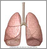
NORMAL
 ABNORMAL
SYMPTOMS
* Depends on the size of p.
* Chest pain:
1. Sudden
2. Sharp or stabbing
3. Chest tightness
4. Made worse by breathing or coughing
* Shortness of breath
* Cyanosis or bluish color to the skin and lips due to hypoxia (low oxygen level)
* Rapid rate of breathing
* Fatigue
* Wheezing
* Anxiety
* Patient may be in Shock or unconscious, especially with trauma or tension p.
CAUSE
* Primary p. -- in healthy individuals this occurs without any underlying diseases
* Secondary -- complication of underlying lung disease
* Spontaneous p. -- often occurs after the rupture of a bulla (a.k.a bleb) or blister (not found in normal lungs), which are small air-filled sacs in the lungs of individuals with certain risk factors. Could be primary or secondary.
HOW THE DIAGNOSIS IS MADE
* History:
1. Symptoms
2. Injuries
3. Procedures or surgeries
4. Illnesses
5. Medications
6. Habits
7. Occupation
8. Allergies
* Medical exam:
1. Skin may show the site of trauma
2. Low Blood Pressure
3. Fever may be present
4. Cyanosis
5. Air can leak under the skin (subcutaneous Emphysema) and has a spongy feel
6. Pallor -- pale skin
7. Rapid heart and respiration rate
8. The chest movements are not symmetrical
9. The trachea (major airway -- located in the center of the neck) may be shifted in tension p.
10. When the doctor listens to the heart or the lungs, the normal sounds may be diminished or absent.
* Tests:
1. Chest X-Ray will show the abnormal air pocket, and Collapsed Lung or structures that have been pushed to one side.
2. CAT scan, using computer imaging, shows detailed views of the chest and lungs.
3. Blood tests may show low oxygen levels (<80 acidosis =" pH">30%:
1. Objective is to remove the air.
2. General physician may consult with a surgeon or a pulmonologist (lung doctor).
3. A needle may be used to remove the air.
4. Aspiration involves inserting a catheter (Teflon tube) into the chest cavity (between the ribs) and attaching to a simple bottle (vacuum inside with no air). This may be enough to expand the Collapsed Lung.
5. A plastic chest tube is inserted through an incision in the chest (between the ribs) and then connected to suction. This method may take few days to drain the trapped air and expand the Collapsed Lung.
* Multiple episodes of Pneumothorax may require surgery or injection of medicines (talc or Doxycycline) into the pleural space.
IF YOU SUSPECT THIS CONDITION
* Contact 911 and seek immediate medical attention. If you've had previous episodes, Quit Smoking and talk to your doctor before high altitude climbing, flying, or scuba diving.
SIMILAR CONDITIONS
* Pericarditis -- inflammation of the sac covering the heart
* Pleurisy -- inflammation of the pleura
* Pulmonary Embolism -- blood clot in the lungs
* Myocardial Infarction -- heart attack


No comments:
Post a Comment